BOOK APPOINTMENT
BOOK APPOINTMENT

Back Surgery

Minimally Invasive Lumbar Microdiscectomy with Foraminotomy

Minimally Invasive Lumbar Microdiscectomy with Foraminotomy is a surgical procedure designed to address issues in the lumbar spine, specifically targeting herniated discs and neural foramen (bony openings through which spinal nerves exit). Here are key points about this procedure:
1. Objective: The primary goal is to relieve pressure on spinal nerves, particularly when a herniated disc is compressing nerve roots as they exit the spinal canal through the neural foramen.
2. Minimally Invasive Approach: Similar to other minimally invasive techniques, this procedure involves small incisions and the use of specialized instruments and microscopic visualization. The surgeon employs magnification tools to precisely target and address the affected disc and neural foramen.
3. Microdiscectomy: The surgeon removes the portion of the herniated disc that is causing compression on the spinal nerves. This is done with microsurgical instruments, minimizing disruption to surrounding healthy tissue.
4. Foraminotomy: In addition to addressing the disc herniation, foraminotomy involves enlarging the neural foramen to ensure there is enough space for the nerve roots to exit without compression. This can be especially beneficial if there are bone spurs or other obstructions contributing to nerve impingement.
5. Indications: This procedure is typically recommended for patients with lumbar radiculopathy caused by a herniated disc and associated neural compression in the lumbar spine.
6. Advantages: Minimally invasive lumbar microdiscectomy with foraminotomy offers advantages such as smaller incisions, reduced postoperative pain, quicker recovery, and less disruption to surrounding tissues compared to traditional open surgeries.
7. Recovery: Patients generally experience a faster recovery with this minimally invasive approach. Patients undergoing this procedure go home the same day. Postoperative care may involve a short period of rest, followed by a gradual return to normal activities. Physical therapy may be recommended for rehabilitation.
As with any medical procedure, the decision to undergo minimally invasive lumbar microdiscectomy with foraminotomy would be based on a thorough evaluation of your condition by a Spine Capital provider.
Intraoperative images of patient undergoing L4/5 minimally invasive lumbar microdiskectomy

Endoscopic Lumbar Discectomy

Endoscopic Lumbar Discectomy is a minimally invasive surgical procedure used to treat herniated or bulging discs in the lumbar spine. Here are key points about this procedure:
1. Minimally Invasive Approach: Endoscopic lumbar discectomy is performed through small incisions using an endoscope, a thin, flexible tube with a camera and light source. This allows the surgeon to visualize and address the affected disc without the need for larger incisions.
2. Visualization: The endoscope provides a magnified view of the surgical site on a monitor, enabling the surgeon to navigate and perform the procedure with precision. This minimizes the disruption to surrounding tissues.
3. Disc Access: The surgeon inserts the endoscope through a small incision, and other specialized instruments are used to remove or shrink the portion of the herniated disc that is compressing the spinal nerves.
4. Indications: Endoscopic lumbar discectomy is typically recommended for patients with a single-level lumbar herniated disc causing symptoms such as lower back pain, leg pain, numbness, or weakness that hasn’t responded to conservative treatments.
5. Advantages: The minimally invasive nature of this procedure often results in less postoperative pain, reduced scarring, quicker recovery, and shorter hospital stays compared to traditional open discectomy procedures.
6. Outpatient Procedure: In many cases, endoscopic lumbar discectomy can be performed on an outpatient basis, allowing patients to go home the same day.
7. Recovery: Recovery time is generally faster compared to traditional open surgeries. Patients may be advised to gradually resume normal activities, and physical therapy may be recommended to aid in rehabilitation.
As with any medical procedure, the decision to undergo endoscopic lumbar discectomy should be based on a thorough evaluation by your Spine Capital provider, taking into consideration the specific condition and medical history of the patient.

Lumbar laminectomy

Lumbar laminectomy, also known as decompressive laminectomy, is a surgical procedure performed to relieve pressure on the spinal nerves in the lumbar (lower) region of the spine. Here are key points about this procedure:
1. Objective: The primary goal of lumbar laminectomy is to alleviate symptoms caused by spinal stenosis, herniated discs, or other conditions that lead to compression of the spinal nerves. The procedure involves removing a portion of the lamina, which is the bony arch on the vertebra, to create more space for the nerves.
2. Surgical Approach: Laminectomy is typically performed through a small incision in the back over the affected vertebrae. The surgeon removes the lamina, and if necessary, other structures like bone spurs or disc material contributing to nerve compression.
3. Indications: Lumbar laminectomy is often recommended for patients experiencing symptoms such as leg pain, numbness, or weakness due to pressure on the spinal nerves. It is commonly associated with conditions like spinal stenosis, herniated discs, or degenerative disc disease.
4. Recovery: Recovery time varies, but patients typically need a period of rest and rehabilitation. Physical therapy may be recommended to help restore strength and flexibility.
The decision to undergo lumbar laminectomy should be made in consultation with a Spine Capital provider, considering your medical history, the severity of symptoms, and the potential benefits and risks of the procedure.
Watch Now

Lumbar laminectomy and fusion

Lumbar laminectomy and fusion is a surgical procedure that combines the removal of part of the lamina (laminectomy) with spinal fusion to address issues in the lumbar (lower) spine. Here are key points about this procedure:
1. Objective: The primary goal of lumbar laminectomy and fusion is to decompress the spinal nerves by removing a portion of the lamina while also providing stability to the spine through fusion. This creates space for nerves and eliminates motion between vertebrae.
2. Indications: Lumbar laminectomy and fusion are typically recommended for conditions such as spinal stenosis, degenerative disc disease, or recurrent disc herniation when there is instability in the spine.
3. Surgical Approach: The procedure is often performed through an incision in the back. The surgeon removes the lamina and any other structures contributing to nerve compression. Bone grafts are then placed in the space created by the laminectomy to promote fusion. Metal hardware such as screws and rods may be used to stabilize the spine.
4. Spinal Fusion: Fusion aims to eliminate motion between the vertebrae, preventing further compression of nerves and promoting stability. The bone grafts stimulate the growth of new bone that fuses the vertebrae over time.
5. Recovery: Recovery from lumbar laminectomy and fusion involves a period of rest and rehabilitation. Patients require a rigid back brace that is to be worn for 12 weeks when upright. Physical therapy may be recommended to help restore strength and flexibility after the 12 week healing period.
Lumbar laminectomy and fusion is a comprehensive approach to addressing both spinal decompression and instability. The decision to undergo this procedure should be based on individual factors, the specific condition, and the recommendations of your Spine Capital provider.
MRI of a patient with debilitating low back and leg pain due to severe lumbar stenosis at L3/4 and L4/5 and spondylolisthesis at L4/5. Patient underwent lumbar laminectomy and fusion (post-operative x-ray on the right) and had resolution of her symptoms.

Anterior Lumbar Interbody Fusion (ALIF)

Anterior Lumbar Interbody Fusion (ALIF) is a surgical procedure used to address spinal issues in the lumbar (lower) spine. Here are key points about ALIF:
1. Objective: The primary goal of ALIF is to stabilize the spine and promote fusion by removing a damaged disc from the front (anterior) and inserting a bone graft or implant in the intervertebral space.
2. Surgical Approach: ALIF is performed through an incision in the abdomen, allowing access to the front of the spine. This approach provides direct access to the lumbar discs without disrupting the spinal muscles from the back.
3. Disc Removal and Fusion: The surgeon removes the damaged disc and inserts a bone graft or a cage filled with bone graft material into the intervertebral space. This promotes bone growth and fusion between the adjacent vertebrae. Titanium screws may be used to anchor the cage and additional screws may be inserted from a very small incision in the back to stabilize the spine and promote a fusion.
4. Indications: ALIF is often recommended for conditions such as degenerative disc disease, disc herniation, or spinal instability that requires stabilization and fusion.
5. Recovery: Recovery after ALIF varies, and patients typically undergo a period of rest and rehabilitation. Physical therapy may be recommended to aid in the recovery process. Patients need to wear a rigid lumbar brace to aid with healing after an ALIF procedure.
ALIF is one of several approaches to lumbar spinal fusion, and the choice of procedure depends on individual factors, including the specific condition, spinal anatomy, and surgeon’s recommendations.
Watch Now
X-ray above is of a patient who had severe low back and leg pain from a pars fracture leading to a condition called isthmic spondylolisthesis. He underwent ALIF with posterior percutaneous screws fixation (post-op x ray shown) with complete resolution of his symptoms

Minimally Invasive Transforaminal Lumbar Interbody Fusion (MIS TLIF)

Minimally Invasive Transforaminal Lumbar Interbody Fusion (MIS TLIF) is a surgical procedure used to treat conditions affecting the lumbar spine, such as degenerative disc disease, disc herniation, or spinal instability. Here are key points about MIS TLIF:
1. Objective: The primary goal of MIS TLIF is to stabilize the lumbar spine and alleviate symptoms by fusing two vertebrae together using a bone graft or synthetic material.
2. Minimally Invasive Approach: Unlike traditional open TLIF, MIS TLIF is performed through smaller incisions. Specialized instruments and imaging techniques, such as fluoroscopy or intraoperative navigation, are used for precise visualization and reduced tissue disruption
3. Surgical Technique: The surgeon approaches the spine from the back, entering through a small incision. The damaged disc is removed, and a bone graft or cage filled with bone graft material is placed in the intervertebral space to promote fusion. Additional hardware, such as screws and rods, may be used to stabilize the spine.
4. Indications: MIS TLIF is often recommended for patients with conditions such as lumbar disc herniation, degenerative disc disease, or spondylolisthesis that require stabilization and fusion.
5. Advantages: The minimally invasive nature of the procedure typically results in less postoperative pain, reduced blood loss, quicker recovery, and shorter hospital stays compared to traditional open TLIF.
6. Recovery: Recovery after MIS TLIF involves a period of rest and rehabilitation. Physical therapy may be recommended to help patients regain strength, flexibility, and mobility.
MIS TLIF is considered an effective approach for achieving spinal fusion with less disruption to surrounding tissues, making it a favorable option for certain patients. The decision to undergo this procedure should be based on individual factors and the recommendations of your Spine Capital provider.
Watch Now
This is the postoperative x-rays of a woman who had severe low back and leg pain due o recurrent disc herniations at L5/S1. She underwent an MIS TLIF procedure with complete resolutions of her symptoms.

Extreme Lateral Interbody Fusion

Extreme Lateral Interbody Fusion (XLIF) is a surgical procedure used to treat conditions affecting the lumbar spine. Here are key points about XLIF:
1. Objective: The primary goal of XLIF is to stabilize the lumbar spine and alleviate symptoms by fusing two vertebrae together using a bone graft or synthetic material.
2. Surgical Approach: XLIF is performed through a minimally invasive lateral approach, meaning the surgery is done from the side. This approach allows the surgeon to access the spine without disrupting the muscles and tissues in the back.
3. Disc Removal and Fusion: The surgeon removes the damaged disc from between two vertebrae and inserts a bone graft or a cage filled with bone graft material. This promotes spinal fusion, which stabilizes the spine over time.
4. Indications: XLIF is often recommended for patients with conditions such as lumbar disc herniation, degenerative disc disease, or spinal instability that requires stabilization and fusion.
5. Advantages: The lateral approach in XLIF minimizes disruption to the muscles and tissues in the back, resulting in potentially less postoperative pain, reduced blood loss, quicker recovery, and shorter hospital stays compared to traditional fusion procedures.
6. Recovery: Recovery after XLIF involves a period of rest and rehabilitation. After XLIF surgery, patients wear a rigid lumbar brace for a period of 12 weeks when out of bed. Physical therapy may be recommended to help patients regain strength, flexibility, and mobility.
XLIF is considered a minimally invasive option for achieving spinal fusion, and its specific advantages may make it suitable for certain patients with lumbar spine conditions. The decision to undergo this procedure should be based on individual factors and the recommendations of your Spine Capital provider.
Watch Now
Images of a patient who underwent L4/5 XLIF for severe lumbar spondylosis and disc collapse causing central and foraminal stenosis causing debilitating low back and leg pain. Post-operative MRI demonstrates indirect decompression using the XLIF technique.

Robotic spine surgery

Robotic spine surgery refers to the use of robotic systems to assist surgeons in performing various spinal procedures. Here are key points about robotic spine surgery:
1. Robotic Assistance: In robotic spine surgery, a surgical robot is used to enhance the precision, control, and visualization of the surgical procedure. The robot is not autonomous but is controlled by the surgeon
2. Preoperative Planning: Robotic systems often include advanced imaging and planning capabilities. Surgeons can use these tools to create a detailed preoperative plan, allowing for more accurate placement of implants or instrumentation.
3. Navigation and Guidance: During the surgery, the robotic system provides real-time navigation and guidance. This can enhance the accuracy of instrument placement, such as screws, and help ensure that the surgeon follows the planned trajectory.
4. Minimally Invasive Procedures: Robotic technology is frequently employed in minimally invasive spine surgery, where smaller incisions are made. The robot assists in navigating through complex anatomy while minimizing disruption to surrounding tissues.
5. Benefits: The potential benefits of robotic spine surgery include increased accuracy, reduced blood loss, shorter hospital stays, faster recovery times, and potentially less postoperative pain compared to traditional approaches.
6. Applications: Robotic systems can be used in various spinal procedures, including spinal fusions, discectomies, and decompressions. They are often utilized in complex cases or when a high degree of precision is required.
It’s important for individuals considering robotic spine surgery to discuss the benefits, risks, and appropriateness of the procedure with their Spine Capital provider and the surgical team. The decision to use robotic assistance is typically based on the specific case and the surgeon’s judgment.

Lumbar Artificial Disk Replacement

Lumbar arthroplasty, also known as total disc replacement or artificial disc replacement, is a surgical procedure designed to treat specific conditions affecting the lumbar (lower) spine, such as degenerative disc disease. Here are key points about lumbar arthroplasty:
1. Objective: The primary goal of lumbar arthroplasty is to replace a damaged or degenerated intervertebral disc with an artificial disc implant. This aims to maintain spinal motion and flexibility while alleviating pain and improving function.
2. Surgical Approach: Lumbar arthroplasty is typically performed through an anterior approach, meaning the surgeon accesses the spine from the front of the body. This allows direct access to the affected disc without disrupting the posterior spinal structures.
3. Implant Placement: The damaged disc is removed, and an artificial disc implant is inserted into the intervertebral space. This implant is designed to mimic the natural function of a healthy disc, allowing for controlled motion.
4. Indications: Lumbar arthroplasty is often recommended for individuals with symptomatic degenerative disc disease who have not responded to conservative treatments. It may be considered when only one or two lumbar discs are affected.
5. Preservation of Motion: Unlike spinal fusion procedures, which aim to restrict motion between vertebrae, lumbar arthroplasty is designed to preserve some degree of spinal motion. This can potentially reduce stress on adjacent discs and joints.
6. Recovery: Recovery after lumbar arthroplasty varies, and patients may undergo a period of rest and rehabilitation. Physical therapy is often included to help restore strength and mobility.
7. Candidates: Not all individuals with lumbar disc issues are suitable candidates for arthroplasty. The decision to undergo this procedure is based on factors such as the specific condition, spinal anatomy, and the overall health of the patient.
It’s crucial for individuals considering lumbar arthroplasty to have a comprehensive discussion with your Spine Capital provider to understand the potential benefits, risks, and expected outcomes based on their specific condition. The suitability for this procedure is determined on a case-by-case basis.

Spinal cord stimulator (SCS) placement

Spinal cord stimulator (SCS) placement is a procedure where a device is implanted near the spinal cord to manage chronic pain. It delivers electrical impulses to interrupt pain signals by stimulating the spinal cord. The procedure involves placing electrodes in the epidural space and a generator under the skin. It's often considered for those with chronic pain unresponsive to other treatments. Consultation with a Spine Capital provider is crucial to determine if SCS placement is suitable for your specific condition.

Kyphoplasty/Vertebroplasty

Kyphoplasty/Vertebroplasty is a medical procedure used to treat vertebral compression fractures, often caused by osteoporosis or spinal tumors. During kyphoplasty, a surgeon uses a balloon to create space within the fractured vertebra and then injects bone cement into the space to stabilize the bone. This can help relieve pain and restore vertebral height. The procedure aims to improve mobility and reduce the risk of further fractures. It's typically considered for individuals with painful vertebral compression fractures. Consultation with your Spine Capital provider is necessary to determine if kyphoplasty or vertebroplasty is appropriate for a specific situation.

Scoliosis Deformity Corrective Surgery

Scoliosis surgery is a medical intervention performed to correct abnormal curvature of the spine, known as scoliosis. The decision to undergo surgery is typically based on the severity of the spinal curvature, the risk of progression, and the impact of scoliosis on the individual's health. Here are key points about scoliosis surgery:
1. Indications: Surgery is often considered for individuals with moderate to severe scoliosis, particularly if the curvature is progressing, causing pain, or affecting lung and heart function.
2. Surgical Techniques:

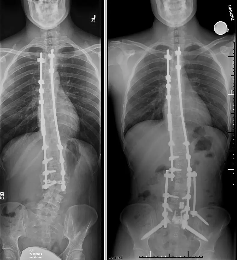
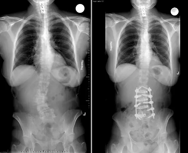
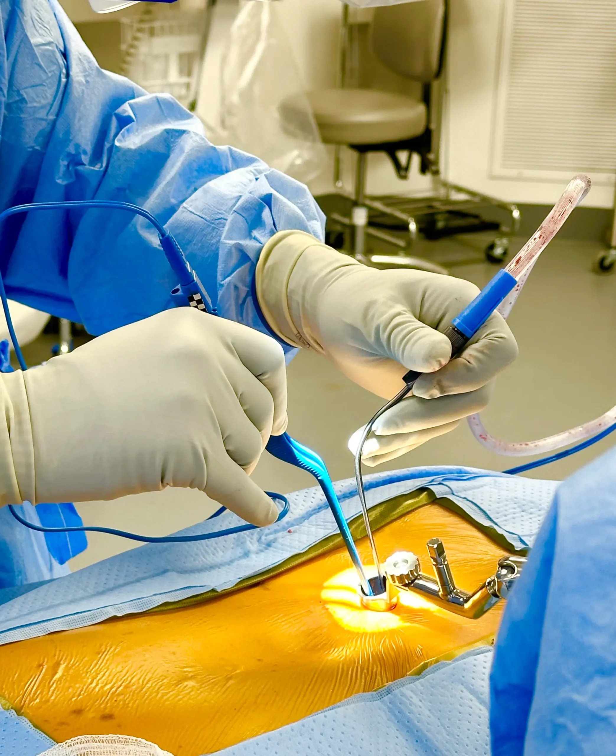
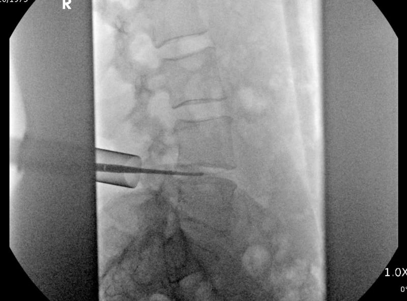
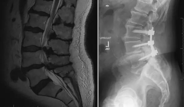
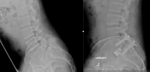
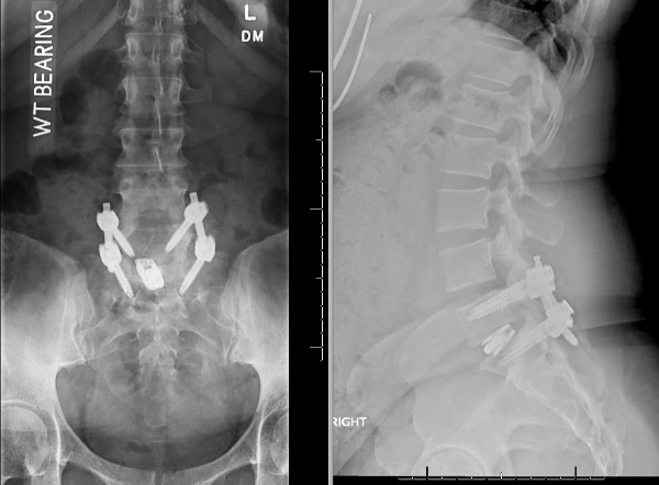
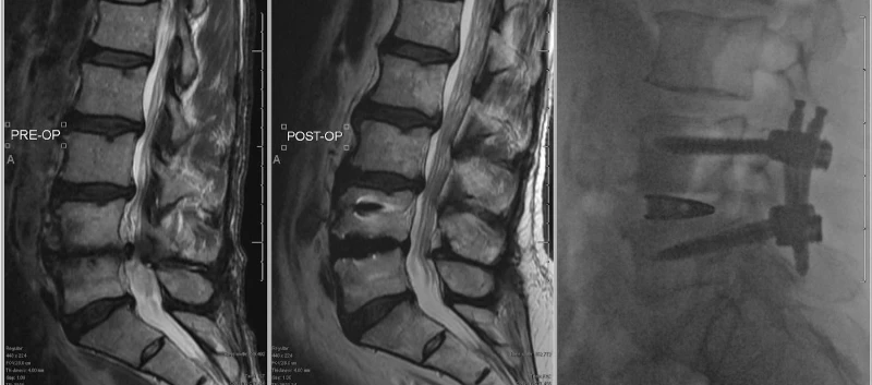
- Spinal Fusion: The most common surgical procedure for scoliosis involves fusing together the vertebrae to stabilize the spine. This is often done using bone grafts, rods, and screws.

 - Instrumentation: Metal rods and screws may be used to correct and maintain the alignment of the spine during the fusion process.

3. Approaches:

  - Posterior Approach: Surgery is performed through the back.

  - Anterior Approach: Surgery is performed through the front of the body.

  - Combined Approach: A combination of anterior and posterior approaches may be used for certain cases.

4. Recovery: Recovery after scoliosis surgery involves a period of rest and rehabilitation. Patients are typically required to wear a brace for some time after surgery. Physical therapy is often a crucial part of the recovery process, helping patients regain strength, flexibility, and proper posture.
The decision to undergo scoliosis surgery is individualized and based on factors such as the degree of curvature, age, overall health, and the impact of scoliosis on the patient's quality of life. It's important for individuals considering surgery to have thorough discussions with their Spine Capital provider to understand the potential benefits, risks, and expected outcomes based on their specific situation.
X-rays of a patient with adolescence idiopathic scoliosis who was treated decades ago with thoracolumbar fusion and as an adult developed degenerative lumbar scoliosis causing debilitating pain as and subsequently underwent corrective scoliosis surgery with resolution of symptoms.

Minimally invasive scoliosis surgery

Minimally invasive scoliosis surgery is an advanced surgical approach aimed at correcting abnormal curvature of the spine (scoliosis) while minimizing disruption to surrounding tissues. Here are key points about minimally invasive scoliosis surgery:
1. Objective: The primary goal of minimally invasive scoliosis surgery is to correct the spinal curvature using less invasive techniques, reducing blood loss, postoperative pain, and promoting a quicker recovery compared to traditional open surgery.
2. Surgical Techniques:
Small Incisions: Minimally invasive techniques involve smaller incisions compared to traditional open surgery.Combining powerful techniques such as extreme lateral lumbar interbody fusion (XLIF), anterior column release (ACR), and posterior percutaneous pedicle screws fixations, complex deformities can be corrected. These techniques focus on muscle-sparing approaches and aim to minimize disruption to muscles and soft tissues surrounding the spine.
3. Instrumentation:
Pedicle Screws and Rods: Specialized instrumentation, such as pedicle screws and rods, may be used to correct and stabilize the spinal curvature.
Guidance Systems: Surgical navigation or imaging guidance systems may assist in precise placement of screws and other instrumentation
4. Patient Selection: Minimally invasive scoliosis surgery may be suitable for certain types and degrees of scoliosis. The decision is based on factors such as the severity of the curvature, the flexibility of the spine, and the overall health of the patient.
5. Indications:
Adolescent Idiopathic Scoliosis (AIS): Minimally invasive approaches are increasingly used for AIS in adolescents.
Adult Scoliosis: In some cases, minimally invasive techniques may be applied to correct scoliosis in adults.
Minimally invasive scoliosis surgery represents a technological advancement in the treatment of scoliosis, offering potential benefits for eligible patients. The decision to undergo this procedure is made on an individual basis after thorough evaluation and discussion between the patient and the healthcare team.
X-rays of a patient who underwent minimally invasive scoliosis surgery for correction of a degenerative lumbar scoliosis causing debilitating low back pain.
Neck Surgery
Copyright © 2024 Spine Capital. All Rights Reserved.
menu